The human heart is depicted in all kinds of ways. But the specific anatomy of the organ itself is absolutely fascinating and you can refer to this heart chart for a better idea of it. As the main organ in your cardiovascular system, the heart is in charge of circulating your blood and providing your body with oxygen. If you're thinking, “That's the lung's job!” you'd also be right. The oxygen your heart is circulating is contained within the blood that the heart helps to pump throughout your body. The heart processes are controlled by your brain and your nervous system. But let's learn more about the parts of the heart, and exactly what they do in your existence (via Cleveland Clinic).
Refer to this amazing heart chart which shows each part perfectly. Read on as we delve into the functions and interactions between the various valves, veins, and chambers of a beating human heart. We'll discuss the performance of each part, and how it plays into the larger role of the heart in your body. Take a second to watch the video below, and then we'll get to the heart of this article, literally!
The Right Side Anatomy Of The Heart
As we get into the specific anatomy of each side of the heart, let's talk about some interesting heart facts. Did you know that, on average, your heart may beat upwards of 115,000 times every day? While it's beating, it's pumping around 2,000 gallons of blood. Also, how long do you think it would be if you were to stretch out the entirety of the blood vessel system connected to your heart? The answer is beyond 60,000 miles (via Healthline). That's 96560.64 kilometres!
The Brachiocephalic Artery
Starting from the top right (left side of the illustration), you'll find the brachiocephalic artery. This artery is a blood vessel branching off the main aorta of the heart. Other names for this artery include the brachiocephalic trunk and the innominate artery. The job of this piece of a functioning heart organ is to supply oxygen-rich blood to the upper right side of your body. Think your right shoulder, neck, and face! the brachiocephalic artery connects to your right carotid artery and your right subclavian artery.
The Superior Vena Cava
Below the Brachiocephalic Artery, you'll find the superior vena cava. This large vein functions as a thoroughfare for deoxygenated blood. The blood is brought through other parts of the body. It goes into the heart to be reoxygenated and sent out again through the circulatory system. The other aspect of this cardiovascular thoroughfare is the inferior vena cava, which we'll get to momentarily.
The Right Pulmonary Arteries
Next up are the right pulmonary arteries. These are also in charge of carrying blood in need of more oxygen from the right side of your body. From there, it goes into the lungs. And here's where the name is important because “pulmonary” actually refers to anything that interacts with the lungs. So the right pulmonary arteries lead directly to your right lung. This blood vessel plays a big very part in the heart.
The Right Pulmonary Veins
Onto the right pulmonary veins which carry the oxygenated blood out of your lungs and into your heart. These are also blood vessels in the pulmonary circuit that work in tandem with the pulmonary arteries.
As you can see in this heart chart, as in the video, the heart is made up of valves, chambers, and veins. All of this helps it to beat and circulate oxygen-rich blood in the right direction for the system.
©viagraphix.net/Shutterstock.com
The Right Atrium
This is one of the upper chambers out of the four chambers of your heart. The right atrium receives deoxygenated blood that comes from that vena cava thoroughfare and passes it through the system. Next, the blood will go to the right ventricle, which we'll also talk about in a few short paragraphs. The chambers of the heart are involved in the processes of heartbeat and blood flow. They have a huge role in a healthy heart.
The Atrioventricular Tricuspid Valve
Moving onto the atrioventricular tricuspid valve, which is, one of four valves in the anatomy of the heart. The atrioventricular tricuspid valve works to pump blood between areas of the heart. This is the mechanism that ensures blood is going between the right atrium (chamber) to the right ventricle. The valve shuts tightly to prevent blood from flowing backward!
The Chordae Tendineae
The chordae tendineae are fibrous connective tissue that appear chord-like and are referred to as “the heartstrings.” These are attached to the valves and the papillary muscles which are within the ventricles. These chords help to stabilize the closure of the ventricle against any backward flow of blood. Overall, the whole system is in place to ensure that blood, both oxygenated and deoxygenated, is flowing in the right direction throughout the circulatory system.
The Right Ventricle
Finally, we come to the right ventricle, which we've talked about a bit before. The right ventricle is yet another heart chamber and it receives the blood from your tricuspid valve. This is then pumped through the pulmonary valve, into pulmonary arteries, and finally into the lungs. Here it goes through a process of reoxygenation and continues in the cycle, eventually making it back into the left atrium. Read on to learn more as we get into functions on the left side of the heart.
The Inferior Vena Cava
And lastly, for this side of the heart, let's talk about the lower half of the deoxygenated blood thoroughfare. The inferior vena cava carries oxygen-poor blood that's coming from your body below. the diaphragm back into the heart system for reoxygenation and circulation. Keep in mind that it's the superior vena cava that handles the upper part of your body. According to the Cleveland Clinic, the vena cava is the body's biggest vein, with the inferior vena cava specifically measuring around 4 inches long and less than an inch in diameter. If you think about the size of your other veins, that is rather large!
The Left Side Anatomy Of The Heart
Onto the left side of the heart, barring the Aorta which is technically more in the middle. However, for the purposes of this article, we'll include it in terms of where it's shown in the video illustration. Did you know that giraffes have lopsided hearts? The left ventricle of their heart is actually thicker because the blood flow has to reach all the way up their long neck to oxygenate their brain!
The Left Common Artery
Starting off with the left common artery which is a blood vessel that supplies blood to your face, your neck, and your brain. Located directly next to the brachiocephalic artery, this artery splits into the left subclavian artery which is just below it at the very top of the heart.
The Left Subclavian Artery
Next, we have the left subclavian artery splits off from the left common artery and sends oxygen-rich blood throughout three sections of your body. It works in tandem with the left common artery and specifically pumps blood to areas of your chest, thyroid, and head, parts of your neck, and into your biceps, shoulders, triceps, and forearms.
The Aorta
Central to the heart, the aorta is a large blood vessel directly in the middle of the heart organ. Size-wise, the aorta is over a foot in length and around an inch across at certain points in its journey across the body. The aorta delivers nutrients, and hormones, to the substances that pass through. It is the main vessel for blood transfer across the heart organ as a whole.
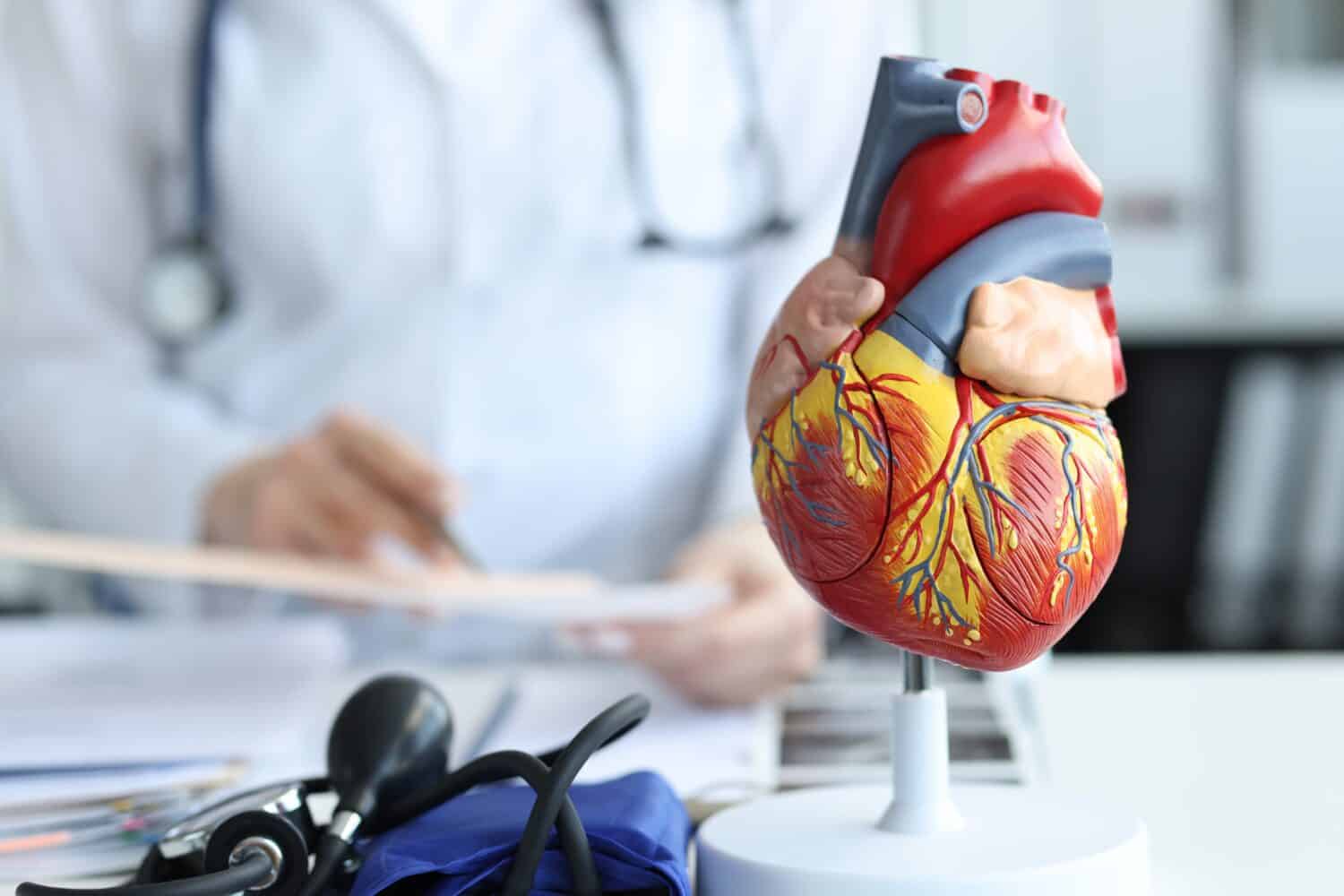
You can see in this heart model, the aorta (the red) is the main heart vessel in the circulatory system.
©H_Ko/Shutterstock.com
The Left Pulmonary Arteries
On the left side of the body, we'll find the left pulmonary arteries. These and their right counterparts carry oxygen-poor blood back into the system for reoxygenation. The left pulmonary artery is linked directly to your left lung and is a necessary part of the whole functioning system.
The Left Pulmonary Veins
Again, like its right counterpart, the left pulmonary veins take the re-oxygenated blood from the lungs back into the carry the oxygenated blood out of your lungs and into your heart. While the heart is absolutely in charge of the pumping, these veins are a huge reason the system works at all.
The Left Atrium
The next chamber of the heart is the left atrium. While the right atrium holds the deoxygenated blood, the left atrium is the reservoir for the oxygenated blood. From this chamber, the blood is sent through to your left ventricle. But first, it has to pass through a series of valves.
The Semilunar Valves
These valves get their name from their shape, which resembles a crescent moon. The semilunar valves are located between the atrium and the ventricle chambers of the heart. They are right next to the atrioventricular mitral valve, another of the four valves in the heart organ.
The Atrioventricular Mitral Valve
Also known as just the mitral valve, this is located between the left atrium chamber and the left ventricle chamber. Like other valves in the system, the mitral valve is a cog in the process of blood flow and circulation. The valves open and close to ensure everything is going in the right direction. In fact, the heartbeat sound that you can hear with a stethoscope is actually made by the valves opening and closing. A working valve ensures that blood doesn't flow backward into another chamber.
The Left Ventricle
The final chamber, the left ventricle, receives the oxygenated blood from the left atrium. It pumps this oxygen-rich blood through to the aortic valve which sends it along throughout your circulatory system. Every heartbeat is a cycle through this system, which makes those 115,000 heartbeats per day make a bit more sense! The left ventricle is an important final step in the whole system.
The Septum
And finally, the structure that divides the right and the left sides of the heart is known as the septum. This is a kind of muscular tissue that separates the walls of the heart. It is divided into the endocardium, the inner layer, the myocardium, which makes up the muscular middle layer of the septum, and the epicardium outer layer which protects the walls. These layers help the heart to function within the body.
The image featured at the top of this post is ©Ahmet Misirligul/Shutterstock.com